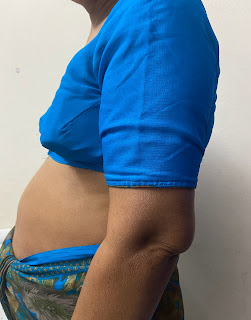
A 36 years old male came with pain abdomen since 4 days
11/12/2023
This is an online E log book to discuss our patient's de-identified health data shared after taking his/her/guardian's signed informed consent.
Here we discuss our individual patient's problems through series of inputs from available global online community of experts with an aim to solve those patient's clinical problems with collective current best evidence based inputs.
This E log book also reflects my patient-centered online learning portfolio and your valuable inputs on the comment.
Sanidha Singh
Roll no.- 98
Unit 4
CHIEF COMPLAINT: Pain abdomen since 4 days
HISTORY OF PRESENTING ILLNESS: Patient was apparently asymptomatic 4 days ago then he developed epigastric pain since 4 days which is dragging type, insidious in onset, gradually progressive and aggravated with food intake and relieved on bending forward.
No h/o fever, cough, breathlessness.
No h/o chest pain, palpitations, orthopnea, paroxysmal nocturnal dyspnoea.
No h/o burning micturition, constipation.
Past History:
N/K/C/O DM, HTN, Asthma, TB, Epilepsy
No h/o CKD, CLD, CAD, CVA
PERSONAL HISTORY:
Diet- mixed
Bowel and bladder- regular
Appetite- normal
Sleep- adequate
Addiction- chronic alcoholic (whiskey : 180 ml daily) since 15 years.
GENERAL PHYSICAL EXAMINATION:
Patient is conscious, coherent,cooperative,well oriented to time,place,person. He is moderately built and nourished.
No signs of pallor, icterus, cyanosis, clubbing, lymphoedenopthy, oedema.
Vitals:
PR- 84 bpm
RR- 16 cpm
BP-140/90mm hg
GRBS- 254 mg/dl
Temp- 98 F
SYSTEMIC EXAMINATION:
CNS: NFND
CVS: S1 S2 +, no murmurs
RS: BAE+
P/A:
Inspection: Abdomen flat, Umbilicus is central and inverted, Appendicectomy scar present, No sinuses, no engorged veins, no visible gastric peristalsis.
Palpating: Abdomen is soft, non distended, tenderness present in the epigastric region, no guarding, no rigidity. No hepatosplenomegaly.
Percussion: liver dullness not obliterated, resonant note heard.
Auscultation: bowel sounds present.
INVESTIGATIONS:
findings
- Elo few hyperechogenic foci noted in the gall bladder all together measuring 9-10 mm
-Elo 32 X18 mm anechogenic cyst noted with internal septations in upper pole of Right kidney
-Elo few cysts noted in left kidney largest @ 12X 11 mm in upper pole.
-Elo altered echotexture of pancreas with calcification and MPD dilated measuring 5 mm and pancreatic cyst- chronic pancreatitis.
-Elo of 34x30 mm heteroechoic lesion with septation noted in left lobe of liver with no internal vascularity — hepatic abscess or hepatic cyst.
-Elo of multiple enlarged lymph nodes along pancreatic region largest measuring 5 mm.
Impression:
- Allered echo texture of pancreas with calcifications & dilated MPD and pancreatic cyst - f/s/o chronic pancreatitis
cholelithialsis - Hepatic cyst or Hepatic abscess in left lobe de liver.
- B/L Renal cysts & Raised echogenicity
- prominent lyphmnodes in para aortic region
Minimal tree fluid in interbowel region & perihepatic region
Review USG done on 11/12/23 i/v/o liquefaction of abscess
E/O 29x30mm and 26X 13mm heteroechoic lesion noted in segment
IV A of the liver with liquefaction of 30-40%
E/O 25 x 21 mm Similar lesion noted in segment IV B of the liver with liquefaction of 40-50%
Impression: liver abscess as described above
CECT SCAN - ABDOMEN & PELVIS done on 12/12/23
IMPRESSION:
Chronic calcific pancreatitis with inflammatory mass in head of pancreas -mass
forming chronic pancreatitis.
Peripancreatic stranding suggests acute inflammation - acute on chronic pancreatitis.
38x32 mm pseudocyst in the lesser sac causing compression of stomach.
Cystic dystrophy of medial wall of 2nd part of duodenum.
Thin fluid collection on the subcapsular aspect of visceral surface of liver.
Few tiny calculi in gall bladders.


Comments
Post a Comment